13

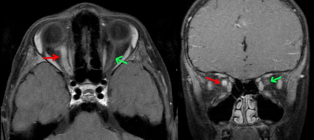
5 year old patient with 1 week of right eye blurry vision, then several days of right eye pain. Physical exam notable for right papilledema and progressively worsening vision.

MRI postcontrast through the orbits shows an enlarged, hyperenhancing right optic nerve (red arrow) compared to the normal left side (green arrow), compatible with optic neuritis.

A lumbar puncture was performed: no oligoclonal bands, no aquaporin 4 IgG, positive anti-MOG. The patient was treated with prednisone with return to normal vision a few months later.

Final diagnosis: optic neuritis from myelin oligodendrocyte glycoprotein​ antibody-associated disease (MOGAD).

11

This patient had episodic electric/shooting/radiating pain of the left face. An MRI was done.

[Top]: Axial heavily T2-weighted image (bright = CSF, dark = not CSF) at the level of trigeminal nerve (cranial nerve V / CN5), shows a normal right CN5 (green). The left CN5 seems to be splayed out by another tubular structure (red), which is the superior cerebellar artery (SCA).

[Bottom Left]: Sagittal reconstruction of the normal right CN5 (along the blue line).

[Bottom Right]: Sagittal reconstruction of the left CN5 shows the left SCA contacting the left CN5. The close proximity of the left SCA and its arterial pulsations likely irritate the cranial nerve, which is the primary sensory nerve of the face, causing trigeminal neuralgia.

6

Incidental finding of a superior lumbar hernia (Grynfeltt-Lesshaft hernia). In this case, only a lobule of retroperitoneal fat is herniating through the defect, but organs can also herniate through.

7

Continuing the theme of things extending into spaces they don't belong in, this is an incidental finding of an inguinal hernia that contains a small portion of the bladder. The patient got the CT for other reasons.

Bowel into inguinal hernia causing bowel obstruction.

Appendix into inguinal hernia, incidental finding.

Ventriculoperitoneal shunt into inguinal hernia, incidental finding.

7

[LEFT]: This patient had one of the longer cerebellar tonsillar herniations I've seen. The tonsil is peg-like in shape and extends quite far below the foramen magnum to the level of the C2 posterior arch. As a result, there is crowding at the foramen magnum that is enough to impede CSF flow, resulting in hydrocephalus with dilated ventricles. Partly seen in the cervical cord from C2 and below is a syrinx, an associated finding. Chiari I is thought to be due to not enough space provided for the cerebellum by the calvarium or skull base shape, causing it to herniate into the spinal canal and cause trouble.

[RIGHT]: A comparison normal from online for you to compare the cerebellar tonsils.

8

[LEFT]: The midbrain has a deep interpeduncular cistern, and the superior cerebellar peduncles are very prominent and elongated, making the brainstem at this level look like a molar tooth. This is a classic finding in Joubert syndrome.

[RIGHT]: A comparison "normal" midbrain. However, this patient's brain is not normal at all. Can you find the abnormalities?

AnswerCompare the left and right temporal lobes in [RIGHT] to the [LEFT] image. Look at how many more gyri and sulci there are in the [LEFT] image. The [RIGHT] patient has a diffuse pachygyria (abnormally reduced brain gyrations). Both Joubert syndrome and pachygyria arise from failure of neurons to migrate, although the genes involved and underlying mechanism are different between the two. (NB: Pachygyria is just a descriptive term for less than normal number of gyri, which can be from a large number of causes mostly having to do with abnormal neuron migration.)

62

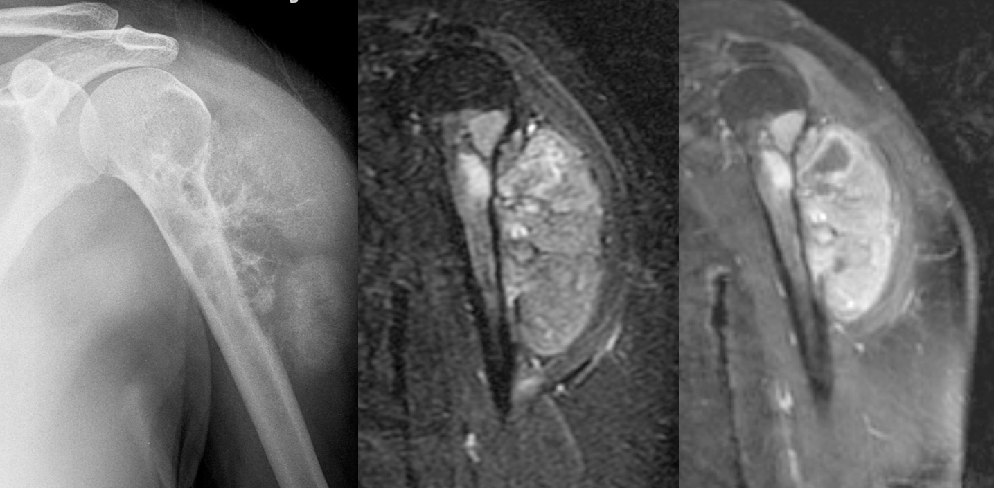
Female in her 30s with painful left shoulder.

[Left]: X-ray shows a mass arising from the left proximal humerus and extending into the adjacent shoulder soft tissues with really aggressive periosteal reaction ("hair on end"). The proximal humerus itself is also heterogeneous with lucent areas. The lateral surface of the upper humerus shows "saucerization," where the cortex is thinned out and looks like a saucer seen on edge.

[Middle]: MRI IR sequence shows a hyperintense bony mass with large soft tissue component.

[Right]: MRI postcontrast T1 IDEAL shows that the mass is enhancing.

This turned out to be high-grade surface osteosarcoma.

8

33 year old female with abdominal pain, abdominal distention, nausea/vomiting, early satiety, and weight loss.

Bottom right: Ultrasound done in a panorama shows how distended the abdomen is by a large multi-cystic mass.

Top right: Non-panoramic ultrasound image shows how limited the imaging modality is in being able to cover such a large mass. This image also shows a more solid area within the mass.

Left: CT images approximately where the ultrasound was done.

The patient underwent laparotomy with removal of the ovarian, fallopian tube, and appendix. There was a large ovarian cyst that was draining serous fluid (watery), mucinous fuid (mucus-like), and blood. The final path was as titled.

14

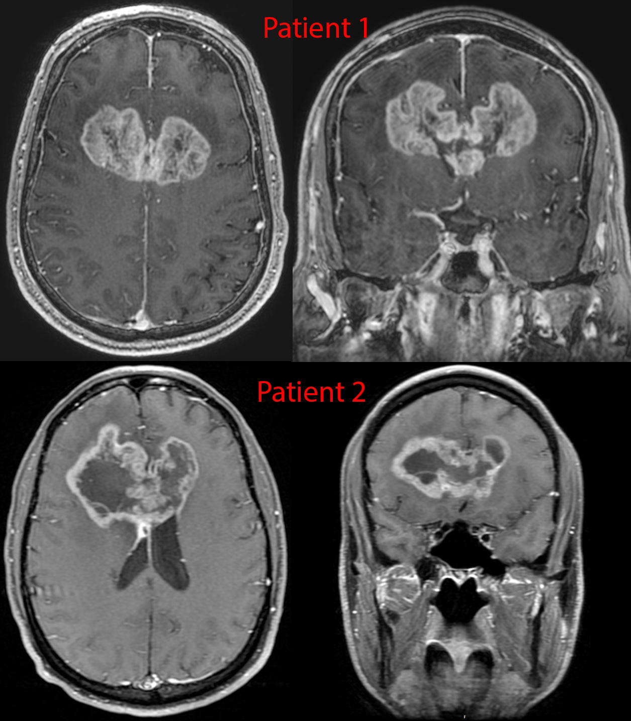
Postcontrast imaging of 2 patients with glioblastoma. These tumors are notorious for spreading along the white matter tracts - in this case the transverse fibers of the corpus callosum, given them a classic "butterfly" appearance.

[-] Spectator@lemmy.world 21 points 1 year ago

I think while the general communities have made it, a lot of niche communities failed to attract enough population to keep on generating more content. As an example, just search for the "Imaginary" series of landscape art communities on the Fediverse (eg. ImaginaryVistas). Many of them don't have any recent posts or 1 post per days or weeks. That's not enough to keep people invested. Even the largest digital art community is still mostly carried by 1 person.

21
submitted 1 year ago* (last edited 1 year ago) by Spectator@lemmy.world to c/radiology@lemmy.world

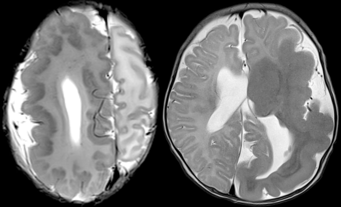
Two different patients with genetic disorders resulting in overgrowth of the brain.

These represent mutations in cell cycle and cell metabolism genes that lead to larger cells and/or more cells. These types of disorders tend to have mosaicism of some form, which is to say some cells have the mutation active while others don't. The distribution of these cells can be very geographic/regional - in these two cases, one hemisphere of the brain is involved.

Compare this against a previous case with hemispheric atrophy.

10
submitted 1 year ago* (last edited 1 year ago) by Spectator@lemmy.world to c/radiology@lemmy.world

Hello everyone!

I am amazed at how quickly this rather specialized community has grown. It gives me perverse pleasure to see that C/Radiology has somehow exceeded C/Medicine in subscriber numbers! So thanks for visiting and allowing me to share my interest in this field with you!

As the community has expanded, we have, of course, come across typical growing pains, and since this is a medical community, some additional factors must also be considered, such as respect for any patient discussions and medical privacy. We have the potential for a lot more growth, but we must be vigilant in respecting medical laws as well. To that end, I have made additional changes to the Community Rules to better clarify the situation for everyone. Additionally, I have conversed with the Lemmy.World admins, who are supportive of this community and now aware of its unique characteristics and requirements.

One major change that has come out of that discussion is that we worry about how inadvertent posts that breach patient confidentiality would behave with federation. It's not like Reddit, where the post is centralized, and there's only one copy to remove. As a consequence, for now, I have changed this community to only allow moderators to post. My hope is that, in the not-too-distant future, Lemmy itself will implement a way for users to post pending moderator approval. Visitors may still comment upon any posts in this Community, and so as a workaround, I've started this megathread for any general questions or discussions you might have regarding radiology. (Please follow the rules still!) If you would like to share a case as a post - please DM me, and I will post on your behalf.

Now onto future updates: For the next few weeks, I will have reduced posting - because I'm going to be away from steady internet. I will continue to post interesting cases I come across thereafter. Eventually, I also plan to have a sticked general guidance on how to look at radiologic images so that you can have a better understanding and capability of looking at these images yourself!

While we're at it, I'm also looking for additional mods to help. I would prefer that you have some medical imaging background, medical background in general, or moderator experience if possible!

14

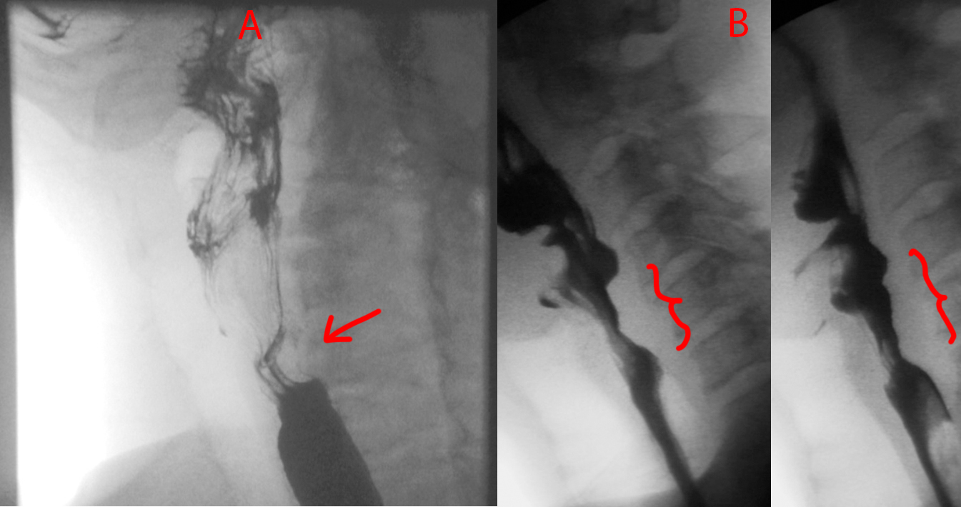
Quick one today. Take a look at Patient A and Patient B.

Patient A has a smooth focal indentation of the posterior cervical esophagus.

Patient B has a broader indentation that is also irregular and nodular along its contour.

Patient A has a cricopharyngeal bar, which is a prominence caused by the cricopharyngeus muscle that can cause dysphagia if it gets really prominent. Patient B has esophageal squamous cell carcinoma.

[-] Spectator@lemmy.world 18 points 1 year ago

Thank you for this comment, because it brings up some very important issues, which I hope this reply addresses.

The biggest issue is the matter of patient confidentiality. This is of utmost concern in an online medical community, especially one wherein clinical vignettes are presented. I take extreme care to avoid including any information that can narrow down to a patient, thus breaching confidentiality. Similarly, I expect anyone else commenting or posting here to follow this rule, Rule 4, which was created not just for internet etiquette but literally to prevent illegal breaches of confidentiality. With regards to consent - this is not required for publishing de-identified information, or sites like Radiopaedia with their thousands of cases would not exist. With regards to patient confidential information that cannot be shared - this not only includes the obvious ones such as patient age, DOB, dates of events, addresses, etc etc, but also vaguer information. For example, there are cases that I would never present here online because the disease is so incredibly rare that the disease itself becomes a patient identifier. These types of cases I would formally publish in the literature if need be. For this particular case, I do not think the information presented breaks these rules (or I would not have posted). Cocaine use is fairly common among the demographic in question, and being found in the shower is not that uncommon, although dramatic.

Second, to address the following:

The title is like your a friend but the text is from a medical professional.

This is something that I have been struggling with. This community is growing at an exceptional rate, and visitors seem to be overwhelmingly from a non-medical background. There are comments that frankly say they do not understand certain things, and other comments and questions imply a lack of experience with looking at imaging studies. I have been vacillating between using terminology and sentences that laypeople can understand versus maintaining medical terminology. I think this is why you think I am writing about a friend in the title, but the body of the post is more medically-oriented. For this reason, I have changed the title - it did not need to be so dramatic. In the future, I will be more careful with my wording.

Please let me know if this addresses your concerns. I would also love to hear more input regarding point #2. Should I continue to word cases as if talking to other medical professionals or include more basic terminology so that the general public can understand? The purpose of these cases, and this community in general, is to be an educational resource in terms of what Radiology is and does.
[-] Spectator@lemmy.world 143 points 1 year ago

Bone break! Why so weak? -> Oh, tumor surprise! -> Tumor growing, bad! Make bone weak(er)! -> Fix fix fix. Luckily not a cancer. -> Good.

[-] Spectator@lemmy.world 30 points 1 year ago

Getting caught in a large rainstorm in the evening while on a mountain trail with 3 more hours of rocky descent to go.

[-] Spectator@lemmy.world 13 points 1 year ago* (last edited 1 year ago)

I was really surprised to hear this, so I dug a little deeper. It looks like an honest mistake.

Here's what their admin said:

We unintionally did until about 8 hours ago.

Lemmy.ml has a ton of bot crawlers in its nginx logs spam fetching posts, so I added the bots to an nginx block. Turns out one called kbinbot wasn’t actually a crawling bot, but their federation requester.

If we don’t respond quickly, its because we have notification backlogs that are months long at this point.

[-] Spectator@lemmy.world 15 points 1 year ago

Just a week and a half ago, we were celebrating going from #2 to #1 most populous instance. And just 2 weeks before that, I had no idea what a lemmy was.

It's like catching a nice surf wave.

[-] Spectator@lemmy.world 16 points 1 year ago

Congrats. I love how every time I log on, there's a new update to wefwe-Voyager.

[-] Spectator@lemmy.world 15 points 1 year ago

It's probably from the lemmy app wefwef. They recently added the ability to report things and the default option is "Spam or Abuse" while the secondary option is to put in a custom text.

[-] Spectator@lemmy.world 79 points 1 year ago

I'm not sure wtf you just said, but lemmy.world feels very smooth today, so thank you for your continued hard work!

[-] Spectator@lemmy.world 22 points 1 year ago

And… we’re back!

[-] Spectator@lemmy.world 45 points 1 year ago

How's that Nazi bar story go again?

I was at a shitty crust punk bar once getting an afterwork beer. One of those shitholes where the bartenders clearly hate you. So the bartender and I were ignoring one another when someone sits next to me and he immediately says, "No. Get out."

And the dude next to me says, "Hey, I'm not doing anything. I'm a paying customer," and the bartender reaches under the counter for a bat or something and says, "Out. Now," and the dude leaves, kind of yelling. And he was dressed in a punk uniform, I noticed.

Anyway, I asked what that was about and the bartender was like, "You didn't see his vest but it was all nazi shit. Iron crosses and stuff. You get to recognize them."

And I was like, "Oh okay," and he continues.

"You have to nip it in the bud immediately. These guys come in and it's always a nice, polite one. And you serve them because you don't want to cause a scene. And then they become a regular and after a while they bring a friend. And that dude is cool too.

And then THEY bring friends, and the friends bring friends, and they stop being cool and then you realize, 'Oh shit. This is a nazi bar now.' And it's too late because they're entrenched and if you try to kick them out, they cause a PROBLEM. So you have to shut them down."

And I was like, "Oh damn," and he said, "Yeah, you have to ignore their reasonable arguments because their end goal is to be terrible, awful people."

And then he went back to ignoring me. But I haven't forgotten that at all.

-iamragesparkle

[-] Spectator@lemmy.world 15 points 1 year ago

I hope at the least that the Beehaw communities are removed from the List of Communities/All page since anyone from this server subscribing to them won't receive any updates. Meanwhile, they are taking up space and preventing other communities from reaching the higher ranks.

view more: next ›

Spectator

joined 1 year ago
MODERATOR OF