1
6
2
8
submitted 4 months ago by Sal@mander.xyz to c/microscopy@mander.xyz
3
1
4
1
submitted 8 months ago by Sal@mander.xyz to c/microscopy@mander.xyz
5
1
6
1
7
2
8
3
9
1
submitted 1 year ago by Sal@mander.xyz to c/microscopy@mander.xyz

A video of a "giant" single-celled organism eating a rotifer!

10
1
11
1
submitted 1 year ago by wjs018@beehaw.org to c/microscopy@mander.xyz

Breakthrough here is the ability to image embryos comprised of living cells as opposed to post-mortem embryos.

original doi: https://doi.org/10.1016/j.cell.2023.06.003

12
1

Next project. :)

13
1

I've gotten all the parts printed and collected but haven't set it up yet. Excited to see how well it does.

14
1
submitted 1 year ago* (last edited 1 year ago) by fossilesque@mander.xyz to c/microscopy@mander.xyz

Tender-handed, stroke a nettle,

And it stings you for your pains.

Grasp it like a man of mettle,

And it soft as silk remains.

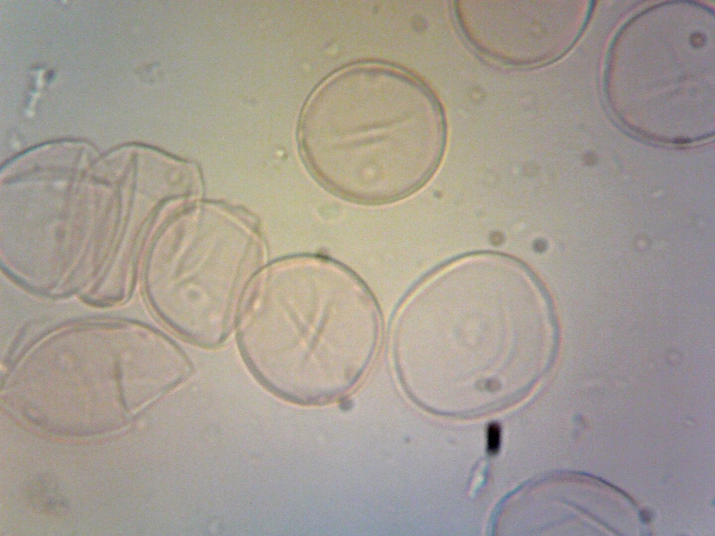
This little guy is Urtica diocia, better known as Stinging Nettle.

It is found all over the world in mild and temperate climates and has a long history of use as a source of traditional medicine, food, tea, and textile raw materials. It's an old treatment for arthritis as its' stings increase blood flow.

In England, it was once thought that the Romans were the first to import the plant. Nettle fibre evidence from a cist on Dartmoor, however, suggests that the plant was collected locally as far back as the Bronze Age.

https://archaeology.co.uk/articles/features/cist-whitehorse-hill.htm

In palaeoecology, it is used as a disturbance indicator, such as on sites of abandoned habitation/construction, within communities of cultivated ground, and on areas enriched with cattle and sheep dung.

Nettles are generally considered to be weeds due to their rapid growth but offer great benefits in rejuvenating over-fertilized soils and increasing local biodiversity.

Personally, I like it in a nice tea. It is surprisingly good. 🍵

Wiki upload: https://commons.wikimedia.org/wiki/File:Urtica_dioica_(Stinging_Nettle)_pollen.tif

I uploaded this a while ago with my beaten up Brunell hobbiest microscope and have since gotten much better ones. Going to resurect this series at some point but here's one for now.

15
1
16
1

Using a technique called high-resolution liquid cell transmission electron microscopy (LC-TEM) at the Molecular Foundry, the researchers captured real-time, atomic-scale LC-TEM videos of Cd-CdCl2 CSNPs ripening in solution.

17
1

Fully solid-state lithium batteries offer some key advantages over the current liquid electrolyte based systems. But these solid electrolytes under development can be unreliable and their degradation mechanisms are unclear. This investigation employed transmission electron microscopy (TEM) to study the evolution of these materials while they operate. They found that differences between the expansion of the cathode material and solid electrolyte induced delamination at their interface. They also noted microscopic cracks forming in the cathode material, and reduction of LCO to metallic Co when the potential was allowed to drop below 1.5 V vs Li/Li+.

18
1
submitted 1 year ago by wjs018@beehaw.org to c/microscopy@mander.xyz

This summarizes a paper in which researchers created a microscope probe using a single, 110 µm diameter optical fiber. An interesting technique from this that I had not seen before, was that the end of the optical fiber is angled and coated with aluminum to create a mirror looking perpendicular to the length of the fiber. This enables researchers to image living cells less intrusively.

doi: https://dx.doi.org/10.1038/s41467-023-36889-z

19
1

Tarantula (Acanthoscurria geniculata) embryo showing expression of genes that pattern the legs and body wall. Right side has curved abdomen, 4 walking legs, and 1 pedipalp (head accidentally removed). Left side has curved abdomen and 1 walking leg (the rest accidentally removed).

The pink/white stripes are where the gene "odd-skipped" is expressed, which marks where the leg joints will form.

20
1

Error message is SyntaxError: Unexpected token 'R', "Request er"... is not valid JSON

Are image uploads not allowed here? Only URLs? Thanks! Hoping this community grows and flourishes! :)

21
1
22
1
submitted 1 year ago* (last edited 1 year ago) by AlchemicalAgent@mander.xyz to c/microscopy@mander.xyz

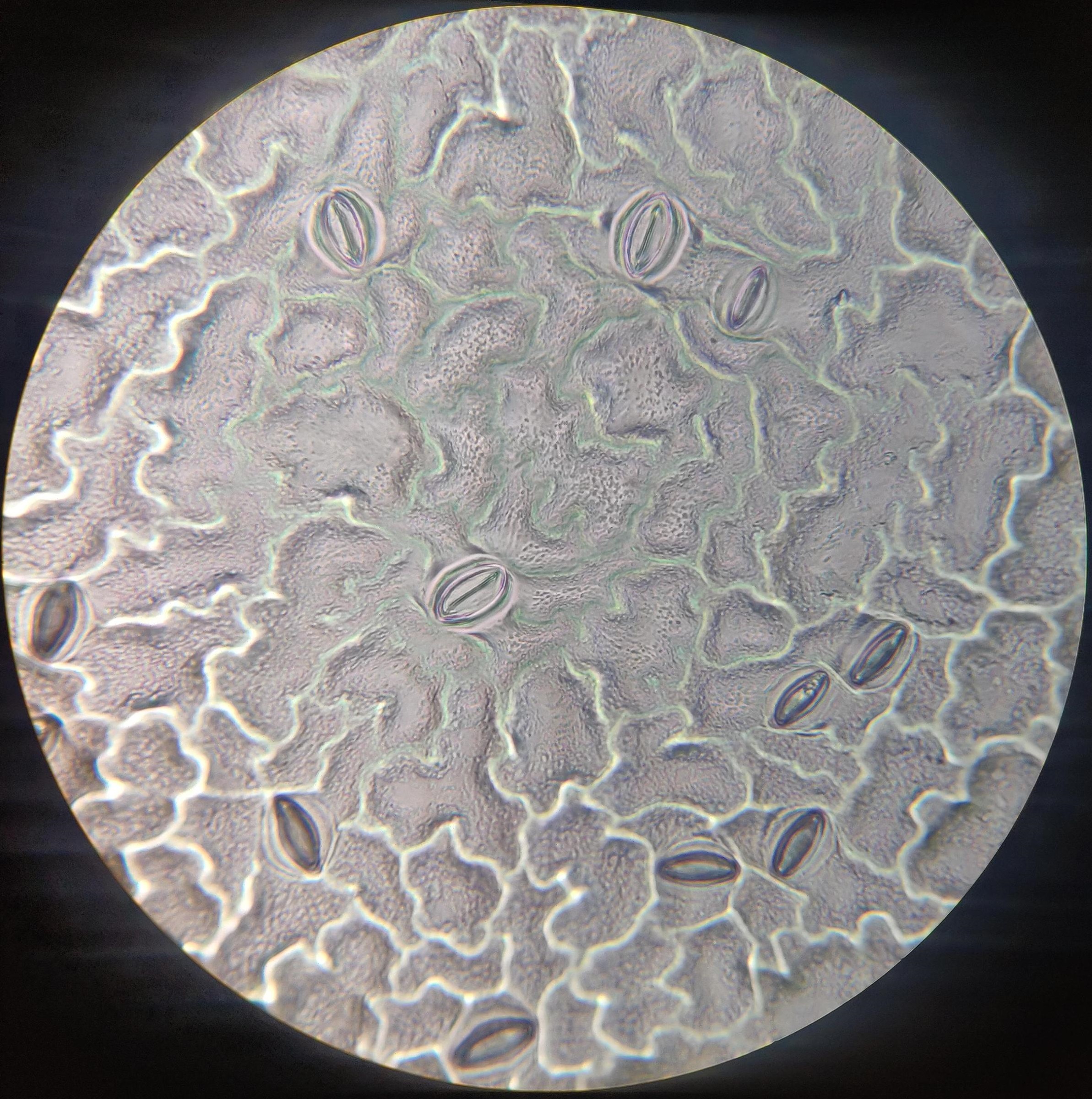
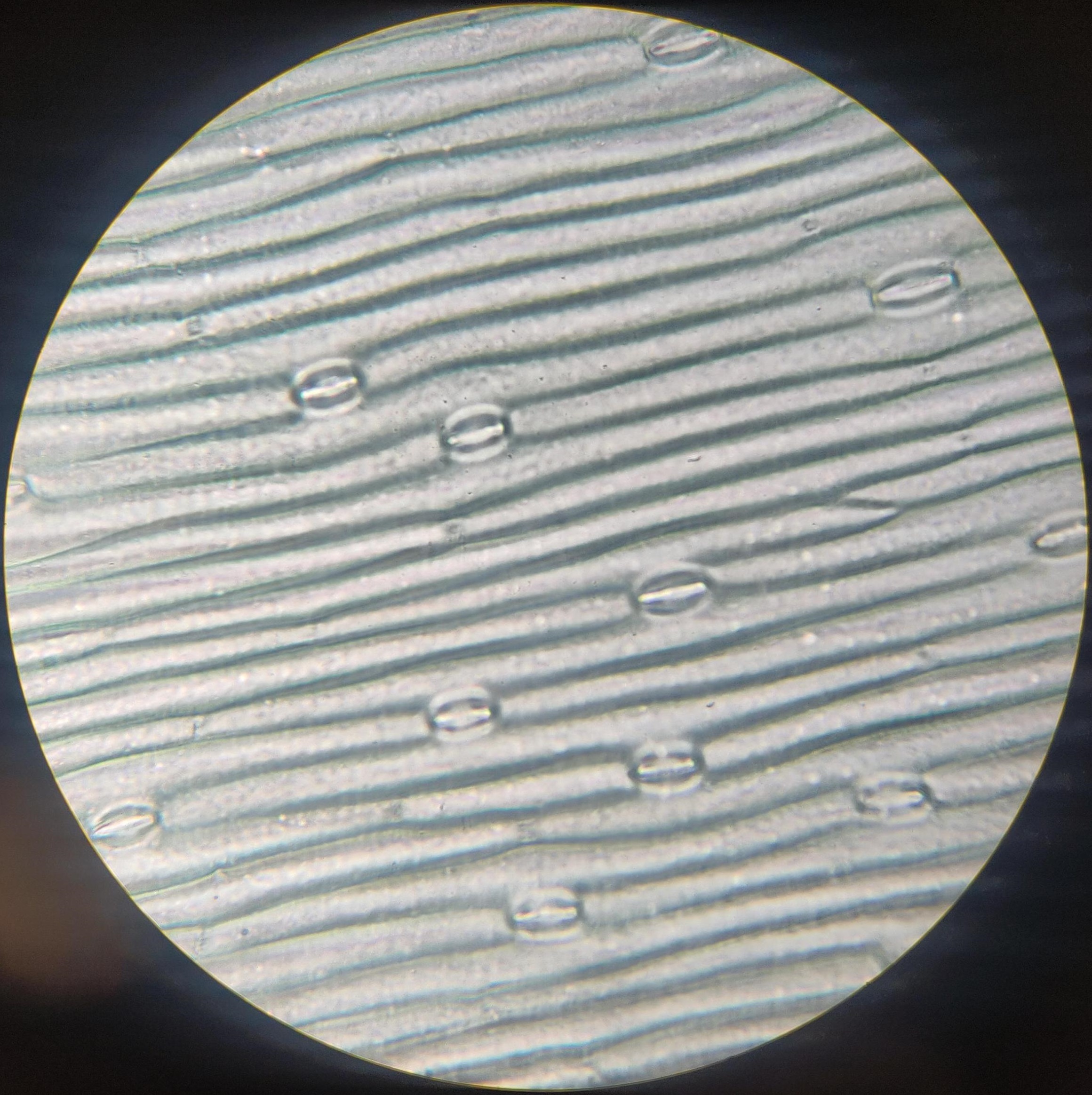
The school year just ended but I wanted to show off what my class was able to accomplish with some cheap high school microscopes. The images below are under-leaf impressions of diocot and monocot plants which show the stomata used to control water evaporation. To avoid the preparation of slicing leaves themselves we painted a thin layer of nail polish on the underside. Peeling this dry layer off results in a mold of the leaf itself which can then be attached to a blank slide with cellophane tape.

Dicot @400x

Monocot @400x

23
1
submitted 2 years ago by Sal@mander.xyz to c/microscopy@mander.xyz
24
1

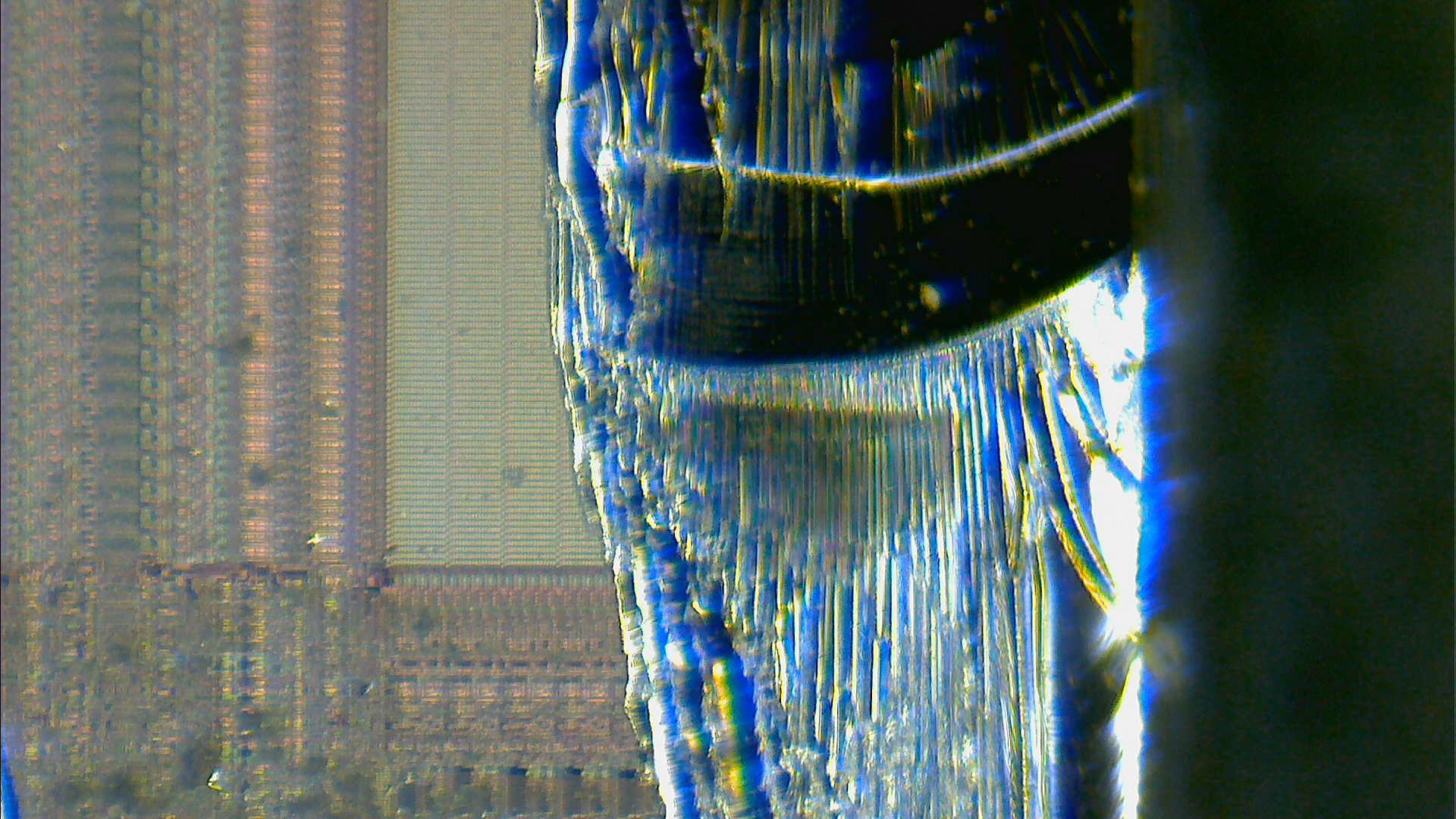
I have to admit, not my most successful attempt at the "apply violence until the die is visible" method of decapping ever. The silver stuff to the right is the broken off silicon.

25
1
submitted 2 years ago by Sal@mander.xyz to c/microscopy@mander.xyz

I put a drop of liquid from my lotus flower's vase under the microscope and noticed a lot of bdelloid rotifers, so I decided to record one at 400x magnification.

I also used this opportunity to upload a video to a PeerTube instance. It is actually a bit of work to find an appropriate instance willing to register new users, as many of them accept a narrow range of topics and 'microscopy videos' is usually not one of them.

view more: next ›

Microscopy

282 readers
1 users here now

Anything related to things that are too small to see them with the eye, and the tools used to observe them.

This space is quite general in scope - microscopes, microbiology, small component electronics, questions about buying optical components, etc.

founded 2 years ago
MODERATORS